






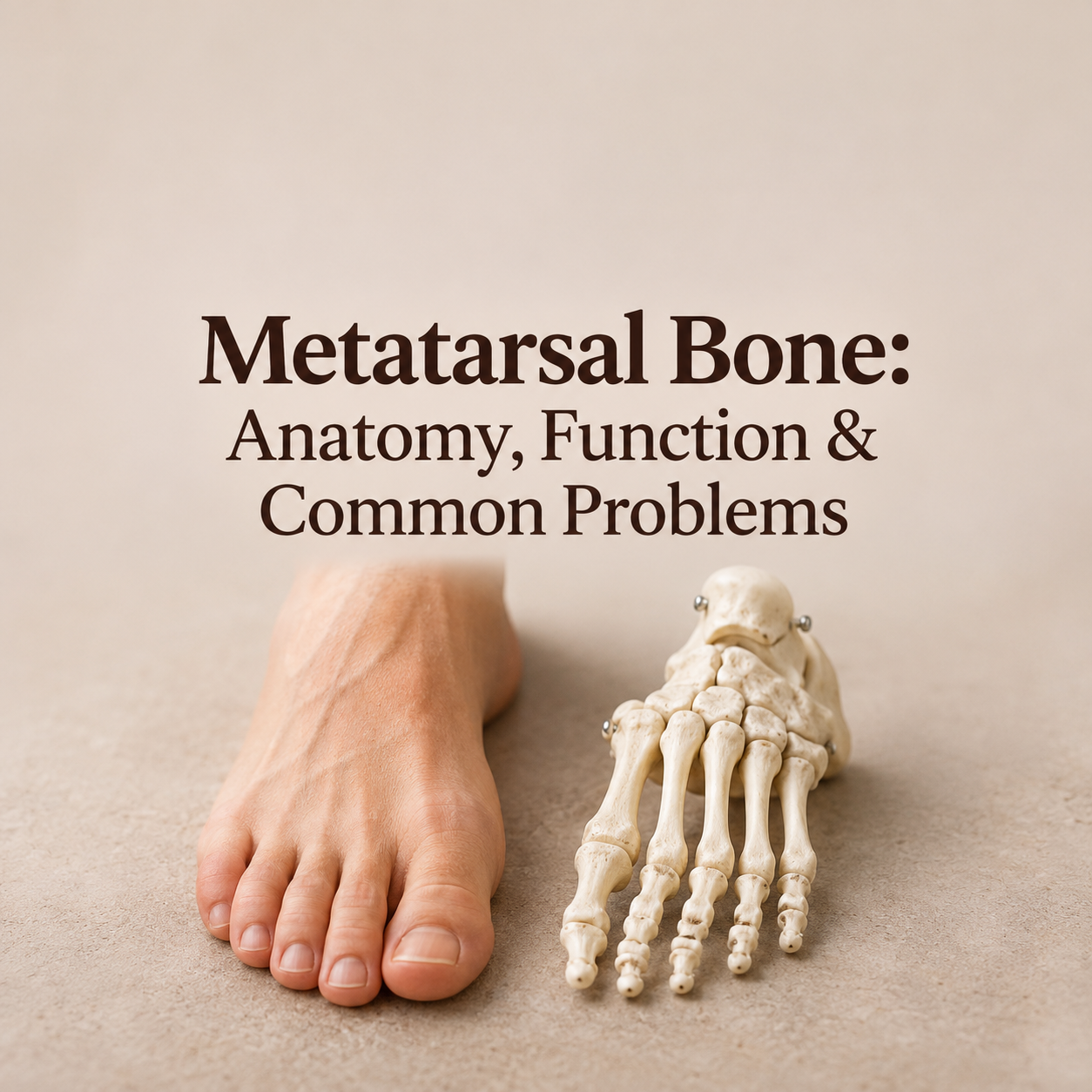
Every step you take, every jump, sprint and tiptoe reach depends on a small group of bones that most people never think about until one of them starts to hurt. These are the metatarsal bones, the five long bones that form the middle of your forefoot and connect your ankle region to your toes. They carry your body weight, help you push off the ground and give your foot the shape it needs to stay balanced on any surface.
In this complete guide you will learn what the metatarsal bone is, where it is located, how it is built, what it does, which injuries and conditions affect it, and what you can do to protect it. Whether you are a student, a runner, a health writer or someone dealing with forefoot pain, this article gives you clear, practical answers.
Quick answer: The metatarsal bones are five long bones (numbered 1 to 5) located between the tarsal bones of the midfoot and the toe bones (phalanges). Their main functions are to bear body weight, act as levers for push-off while walking and running, form the arches of the foot, help with balance and provide attachment points for muscles, tendons and ligaments.
What Is a Metatarsal Bone?
The word metatarsal comes from the Greek meta (beyond) and tarsos (the flat of the foot or ankle region). In simple words, a metatarsal is a bone that lies “beyond the tarsus.” Each foot has five of them, so a person has ten metatarsal bones in total.
The human foot contains 26 bones: 7 tarsal bones, 5 metatarsal bones and 14 phalanges. Together with 33 joints and more than 100 muscles, tendons and ligaments, they make the foot one of the most complex structures in the body. The metatarsals are the longest bones in the foot after the heel bone, and they work as the bridge between the rigid back of the foot and the flexible toes.
Quick Facts at a Glance
| Feature | Details |
|---|---|
| Number | 5 in each foot (10 in total) |
| Names | 1st to 5th, counted from the big-toe side to the little-toe side |
| Location | Forefoot, between the tarsal bones and the toe bones |
| Bone type | Long bones |
| Parts | Base, shaft (body), neck and head |
| Main joints | Tarsometatarsal (TMT), metatarsophalangeal (MTP) and intermetatarsal joints |
| Longest | Usually the 2nd metatarsal |
| Shortest and thickest | The 1st metatarsal |
| Main roles | Weight bearing, push-off, balance, arch support, muscle attachment |
Where Are the Metatarsal Bones Located?
Look at the top of your foot and slide a finger from your ankle toward your toes. The long, slightly ridged bones you feel under the skin in the middle of the foot are your metatarsals. They sit in three layers of the foot’s structure:
- Behind them: the tarsal bones (three cuneiforms, the cuboid and the navicular), which form the midfoot.
- In front of them: the phalanges, the small bones of the toes.
- Beside each other: the neighbouring metatarsals, which are tied together by strong ligaments and small muscles.
The metatarsal heads form the “ball” of your foot, which is the area you press into the ground when you rise onto your toes. This is why pain in this region is so noticeable during walking, running and standing.
Anatomy of a Metatarsal Bone
All five metatarsals share the same basic structure. Each one is divided into four parts:
- Base (proximal end): the wider end closest to the ankle. It forms the tarsometatarsal joints with the tarsal bones and also touches the neighbouring metatarsal bases. These joints are tightly bound by ligaments and give the midfoot its stability.
- Shaft (body): the long middle section. It is slightly curved, which helps the bone absorb forces, and it provides a surface where small muscles called interossei attach.
- Neck: the narrow section just behind the head. Because it is thinner, it can be a weak spot in some injuries.
- Head (distal end): the rounded end that meets the proximal phalanx of each toe to form the metatarsophalangeal (MTP) joint. The head is covered with smooth cartilage so the toe can bend freely.
The Five Metatarsals One by One
Although they look similar, each metatarsal has its own personality and job.
1st Metatarsal (Big Toe Side)
The first metatarsal is the shortest and the thickest of the five. It connects with the medial cuneiform and supports the big toe. Under its head sit two tiny bones called sesamoids, which are embedded in a tendon and act like pulleys, protecting the tendon from pressure. Because the big toe drives the final push of every step, the first metatarsal carries more load than any other. The tendons of tibialis anterior and peroneus longus attach near its base.
2nd Metatarsal
The second metatarsal is usually the longest. Its base sits recessed between the medial and lateral cuneiforms, like a keystone in an arch. A strong band called the Lisfranc ligament anchors this bone to the medial cuneiform, making the second metatarsal the key to midfoot stability. It is also the metatarsal most often affected by stress fractures.
3rd Metatarsal
The third metatarsal joins the lateral cuneiform. It forms the central column of the foot and works closely with the second to share weight during standing and walking.
4th Metatarsal
The fourth metatarsal is slimmer and joins mainly with the cuboid. It acts as a bridge between the central and outer columns of the foot and helps the forefoot adapt to uneven ground.
5th Metatarsal (Little Toe Side)
The fifth metatarsal forms the outer edge of the foot. At its base is a bony bump called the tuberosity, which you can feel on the outside of your foot. The peroneus brevis tendon attaches here, and the peroneus tertius attaches along the top of the base. This bone is well known for the Jones fracture and other injuries.
Joints and Ligaments Around the Metatarsals
Bones do not work alone. The metatarsals are linked by several important joints:
- Tarsometatarsal (TMT or Lisfranc) joints: connect the metatarsal bases to the cuneiforms and cuboid. They allow only small movements but provide great stability.
- Intermetatarsal joints: small joints between the bases of neighbouring metatarsals, reinforced by ligaments.
- Metatarsophalangeal (MTP) joints: connect each metatarsal head to its toe. They bend upward as you push off, which is essential for a smooth walking pattern.
- Plantar plate and deep transverse metatarsal ligament: thick tissue under and between the metatarsal heads that keeps them aligned and supports the ball of the foot.
7 Key Functions of the Metatarsal Bones
1. Weight Bearing
When you stand, your body weight is shared mainly between the heel and the heads of the metatarsals. The first and fifth metatarsal heads, together with the heel, form a “tripod” that keeps you steady. During walking and running, the load shifts forward and the metatarsal heads take a large share of the force with every step.
2. Push-Off and Propulsion
As your heel lifts, your body weight rolls onto the metatarsal heads and toes. The metatarsals act as rigid levers that transfer force from your calf muscles to the ground, driving you forward. This is one reason that even a small injury here can change the way you walk.
3. Forming the Arches of the Foot
The metatarsals are part of the three arches of the foot: the medial longitudinal arch, the lateral longitudinal arch and the transverse arch that runs across the metatarsal heads. These arches act like springs, storing and releasing energy as you move.
4. Shock Absorption
The slight curve of each shaft, the flexible joints and the cushioning fat pad under the metatarsal heads work together to absorb impact when your foot hits the ground. Without this system, more shock would travel up to your ankles, knees and spine.
5. Balance and Stability
The metatarsals widen the base of support of the foot. Their joints and ligaments also send position information to the brain, helping you stay balanced when you stand on one leg, climb stairs or walk on rough terrain.
6. Attachment Points for Muscles and Ligaments
Many muscles and tendons anchor on the metatarsals, including tibialis anterior, peroneus longus, peroneus brevis and peroneus tertius. The small interossei muscles arise from the shafts, and adductor hallucis helps control the big toe. Together they steer the foot, support the arches and fine-tune movement.
7. Protecting Soft Tissues
The bony framework gives protection and a channel for the nerves, blood vessels and tendons that travel to the toes, helping them stay safe during movement and pressure.
How the Metatarsals Work When You Walk
Walking is a smooth sequence in which the metatarsals play a different role at each stage:
- Heel strike: the heel touches the ground first and the metatarsals are mostly passive.
- Mid-stance: the foot flattens slightly and the metatarsal heads share your body weight to keep the foot stable.
- Heel rise: the heel lifts and the weight moves onto the metatarsal heads.
- Toe-off: the big toe bends upward and tightens the plantar fascia (the windlass mechanism), which raises the arch and turns the foot into a stiff lever. The first metatarsal then delivers the final push.
Muscles and Tendons Linked to the Metatarsals
| Muscle | Where it connects | What it does |
|---|---|---|
| Tibialis anterior | Medial cuneiform and base of the 1st metatarsal | Lifts the foot upward and supports the inner arch |
| Peroneus longus | Base of the 1st metatarsal and medial cuneiform | Pulls the first metatarsal down and supports the arch |
| Peroneus brevis | Tuberosity of the 5th metatarsal | Turns the foot outward and steadies the ankle |
| Peroneus tertius | Top of the base of the 5th metatarsal | Helps lift the foot and turn it outward |
| Dorsal and plantar interossei | Shafts of the metatarsals | Stabilise and move the toes |
| Adductor hallucis | Bases of the 2nd to 4th metatarsals and nearby ligaments | Draws the big toe in and supports the transverse arch |
Common Metatarsal Problems
Because the metatarsals take so much repeated load, they are a frequent site of pain and injury. Here are the most common problems.
Metatarsal Fractures
A break can be caused by a direct blow, a twisting injury, a fall or dropping a heavy object on the foot. Metatarsal fractures are among the most common foot fractures. The fifth metatarsal is especially vulnerable, and there are three typical areas of injury.
Metatarsal Stress Fractures
A stress fracture is a tiny crack caused by repeated pressure rather than a single event. It often affects runners, dancers, military recruits and anyone who suddenly increases activity. The second and third metatarsals are the most common sites. Pain usually builds up slowly, gets worse with activity and eases with rest.
Metatarsalgia
Metatarsalgia is a general term for pain and inflammation in the ball of the foot, under the metatarsal heads. It often feels like a burning or aching pain, or like having a pebble under your foot. Common causes include high-impact sports, tight or high-heeled shoes, foot deformities and excess body weight.
Freiberg’s Disease
This is a condition in which the blood supply to a metatarsal head (most often the second) is disturbed, causing the bone to collapse slightly. It mainly appears in teenagers and young adults and causes pain and stiffness at the base of the toe.
Bunions (Hallux Valgus)
In a bunion, the first metatarsal drifts toward the middle of the body while the big toe angles toward the other toes, creating a painful bump at the joint. Narrow shoes and inherited foot shape both play a role.
Sesamoiditis
Irritation or inflammation of the two small sesamoid bones under the first metatarsal head, usually from repeated pressure in running, dancing or wearing high heels.
Lisfranc Injury
An injury to the tarsometatarsal joint area, often involving the ligament that holds the second metatarsal to the medial cuneiform. It can range from a sprain to a fracture-dislocation and sometimes appears as bruising on the sole of the foot after a twisting injury.
Morton’s Neuroma (A Nearby Nerve Problem)
Although it is a nerve problem and not a bone problem, Morton’s neuroma develops between the metatarsal heads, most often between the third and fourth. It causes burning pain, tingling or a feeling of a folded sock under the ball of the foot.
| Condition | What happens | Typical signs |
|---|---|---|
| Fracture | Break in the bone from injury | Sudden pain, swelling, bruising, trouble walking |
| Stress fracture | Small crack from overuse | Gradual pain that worsens with activity |
| Metatarsalgia | Inflamed ball of the foot | Burning or aching pain under the metatarsal heads |
| Freiberg’s disease | Metatarsal head loses blood supply | Pain and stiffness at the base of a toe |
| Bunion | 1st metatarsal shifts out of line | Bump at the base of the big toe, redness, soreness |
| Sesamoiditis | Irritated sesamoid bones | Pain under the big toe joint |
| Lisfranc injury | Midfoot joint or ligament damage | Midfoot swelling, bruising on the sole, pain when bearing weight |
Symptoms You Should Not Ignore
See a doctor or visit urgent care if you notice any of the following:
- Severe pain after an injury, or being unable to put weight on the foot
- Obvious swelling, bruising (especially on the sole) or a changed foot shape
- Numbness, tingling, a cold or pale foot, or open wounds
- Forefoot pain that lasts more than a few days or keeps coming back
- Pain at night or pain at rest
How Metatarsal Problems Are Diagnosed
A doctor will usually start by asking how the pain began and examining your foot, pressing along each metatarsal and watching how you walk. An X-ray is the most common test. Stress fractures may not appear on an early X-ray, so an MRI, bone scan or a repeat X-ray after a couple of weeks may be used. Weight-bearing X-rays are important when a Lisfranc injury is suspected.
Treatment Options
Treatment depends on the type and location of the problem:
- Rest and activity change: stopping the activity that causes pain is the first step for most overuse problems.
- Ice and elevation: help to reduce swelling and discomfort in the early days.
- Protective footwear: a stiff-soled shoe, post-operative shoe or walking boot reduces stress on the bones while they heal.
- Cushioning and support: metatarsal pads and orthotic insoles can shift pressure away from painful areas.
- Medication: over-the-counter pain relievers may help, but check with a doctor or pharmacist first.
- Physical therapy: exercises for the calf, foot and ankle improve strength and movement once healing allows.
- Surgery: sometimes needed for displaced fractures, unstable Lisfranc injuries, some Jones fractures, severe bunions or when other treatments fail.
How Long Does Recovery Take?
Many simple metatarsal fractures and stress fractures heal in about 6 to 8 weeks, although some, such as Jones fractures and severe midfoot injuries, can take longer. Metatarsalgia often improves within weeks once pressure is reduced. Always follow your doctor’s advice before returning to running or sport.
How to Protect Your Metatarsal Bones
- Choose the right shoes: look for a roomy toe box, good forefoot cushioning and a firm heel counter. Limit high heels and very narrow styles.
- Increase activity gradually: add distance or intensity in small steps, especially if you run or train for sport.
- Replace worn shoes: running shoes usually need replacing after roughly 500 to 800 km.
- Strengthen your feet and calves: toe curls, heel raises and balance exercises support the forefoot.
- Look after bone health: eat enough calcium, vitamin D and protein, and do regular weight-bearing exercise. Low energy intake and low bone density raise the risk of stress fractures.
- Maintain a healthy weight: less body weight means less force on every step.
- Vary your surfaces: alternate hard and soft surfaces and cross-train with low-impact exercise like swimming or cycling.
- Listen to early pain: a small ache in the forefoot is a warning sign. Rest early to avoid a longer layoff later.
Foot-Care Products People Commonly Consider
The right support can make daily life more comfortable when the forefoot is sensitive. The categories below are commonly used for metatarsal comfort. Choose based on your own foot shape, activity and your clinician’s advice.
Metatarsal Pads
Small gel or foam domes placed just behind the ball of the foot to spread pressure away from the metatarsal heads. What to look for: medical-grade gel or soft foam, a size that fits your shoe, and a secure adhesive or sleeve.
Cushioned Insoles and Orthotics
Insoles with arch support and forefoot cushioning can improve how weight is shared across the foot. Over-the-counter options suit mild discomfort, while custom orthotics are made from a scan or mould of your foot for specific problems. What to look for: firm arch support, a soft forefoot layer and a design that fits your usual shoes.
Wide Toe Box and Rocker-Sole Shoes
Shoes with a roomy toe area let the metatarsal heads spread naturally, and a rocker sole helps the foot roll forward with less bending at the ball of the foot. What to look for: a thick, stable midsole, a flexible but supportive upper and space for your toes to move.
Stiff-Soled Shoes and Walking Boots
These are usually recommended by a doctor after a fracture or surgery to limit movement while the bone heals. Use them only as advised and make sure the fit is correct.
Toe Spacers and Bunion Sleeves
Soft silicone spacers and cushioned sleeves can ease rubbing and pressure from a bunion or crowded toes. They provide comfort but do not correct the bone alignment.
Cold Packs and Compression Support
Reusable cold packs and light compression sleeves help manage swelling and soreness after activity or in the early stage of an injury.
Frequently Asked Questions
How many metatarsal bones are in the foot?
There are five metatarsal bones in each foot, so ten in the whole body. They are numbered from 1 to 5, starting at the big-toe side.
What is the difference between metatarsals and tarsals?
Tarsals are the seven short, blocky bones of the midfoot and hindfoot, including the heel bone and the ankle bone. Metatarsals are the five long bones in front of them that lead to the toes.
Which metatarsal is the longest?
The second metatarsal is usually the longest, while the first is the shortest but also the thickest and strongest.
Can you walk on a broken metatarsal?
Some people can still walk on a metatarsal fracture, especially a stress fracture, but doing so can make the injury worse. If you suspect a fracture, get it checked and avoid heavy use until a professional gives advice.
What is a Jones fracture?
A Jones fracture is a break in the fifth metatarsal at the junction between the base and the shaft. The area has limited blood supply, so it may heal slowly and sometimes needs surgery.
What causes pain in the ball of the foot?
Common causes include metatarsalgia, stress fractures, Morton’s neuroma, sesamoiditis and poorly fitting shoes. A professional examination can identify the exact cause.
Conclusion
The metatarsal bones may be small compared with the thigh or spine, but they do an enormous job. These five long bones carry your body weight, shape the arches, provide leverage for every step and anchor important muscles and ligaments. Understanding how they are built and what they do helps you spot problems early, choose better footwear and keep your feet strong for life.
If you feel persistent pain in the middle or front of your foot, do not wait for it to go away on its own. Early care protects the metatarsals and helps you stay active and comfortable.
Medical disclaimer: This article is for general education only and is not a substitute for professional medical advice, diagnosis or treatment. Always seek the advice of a qualified healthcare provider about any foot injury or health concern.